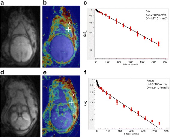
Fig. 4.

Foetal brain. a DW axial images showing the foetal brain at the level of the third ventricle. b Perfusion fraction map at the same level. c IVIM model fitting curve of the foetal brain based on a frontal white matter VOI. d DW axial images showing the foetal brain and the central part of the placenta. e Perfusion fraction map at the same level. f IVIM curve of the central placenta. Cross: central part of the placenta, asterisk: basal plate of the placenta
