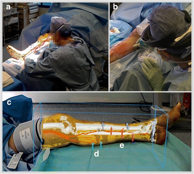
Fig. 3.

a Case 3 AR overlay of models as viewed from remote HoloLens; (b) confirmation of perforator location with audible Doppler ultrasonography. c Case 6 overlay with bounding box; arrows highlighting position of (d) medial sural and (e) posterior tibial perforators
