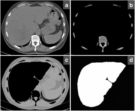
Fig. 2.

Preparation of the patient-inspired in-silico synthetic liver model for the numerical SPCCT experiments. a Original CT image of a healthy liver, (b) segmented bone, (c) segmented soft tissues, and (d) liver part of the original CT image. The liver is removed from the soft tissue image and is replaced in the synthetic liver phantom with a homogenous attenuation of 50 HU
