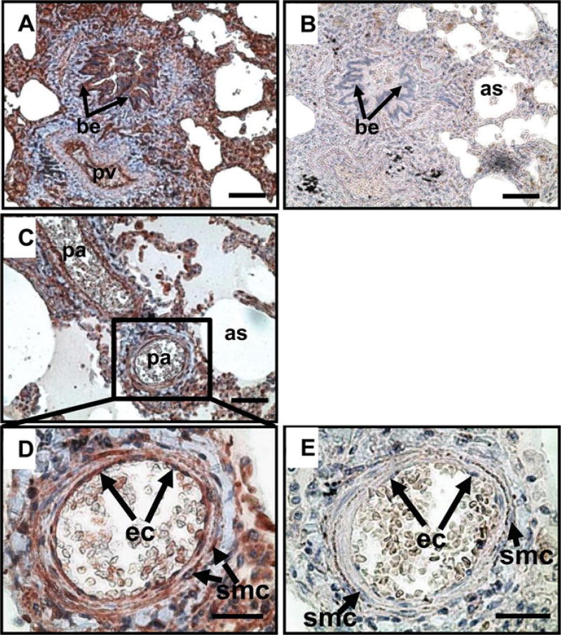
Figure 1.
Immunolocalization of AhR in human lung tissues. A, C, D): The lung tissue microarray was probed with a rabbit antibody against AhR. B, E): The lung tissue microarray was probed with rabbit preimmune IgG, serving as a negative control. Reddish color indicates positive AhR staining. Representative images are shown. be: bronchial epithelium; pv: pulmonary vein; pa: pulmonary artery; as: alveolar sac; ec: endothelial cells. Bars in A-C: 80 µm; Bars in D,E: 40 µm.

