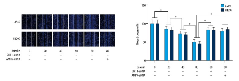
Figure 2.
Effects of baicalin treatment on the wound healing assay results of the non-small cell lung cancer (NSCLC) cells, A549 and H1299. The upper panel of the figure shows the fluorescence images of cultured A549 cells and H1299 cells during the wound healing assay. The nuclei of the cells are positively-stained blue with 4′,6-diamidino-2-phenylindole (DAPI). The columns on the lower panel show the percentage of wound closure involving cultured A549 cells (white columns) and H1299 cells (black columns) treated with baicalin and/or small interfering RNA (siRNA) silencing of the SIRT1 and AMPK genes. * Indicates differences that were statistically significant.

