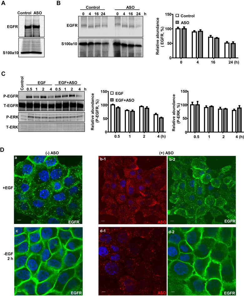
Figure 4.
PS-ASOs do not affect EGFR synthesis, degradation and recycling. (A) A431 cells were pulse labeled with [35S]-Met/Cys for 50 min. EGFR and S100a10 were immunoprecipitated using their specific antibodies, and analyzed by SDS-PAGE. (B) Intracellular levels of nascent EGFR were chased and analyzed by SDS-PAGE at indicated times after the labeling and were visualized and quantified by autoradiography, as in (A). S100a10 was detected and used as a loading control. (C) Protein samples were analyzed by western analyses from A431 cells treated with EGF or EGF and PS-ASOs. The blot was probed sequentially with antibodies specific to the various proteins and the images were quantified by ImageLab (Bio-Rad). (D) Representative images of immunofluorescent staining for EGFR (green) in A431 cells incubated with EGF (a) or EGF and Cy3-labeled PS-ASOs (red) (b-1 and b-2) for 16 h, and then 2 h after the removal of EGF (c) or EGF and Cy3-labeled PS-ASOs (d-1 and d-2). The nuclei were stained with Hoechst 33342 (blue). Scale bars, 5 μm.

