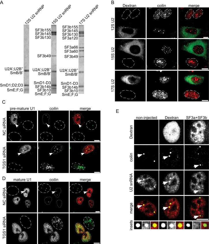
Figure 7.
Partially-assembled snRNP particles induce formation of CBs. (A) Purified 12S U2 snRNP and in vitro reconstituted 15S and 17S U2 snRNPs were analyzed by SDS-PAGE and proteins were visualized by Coomassie staining. (B) TGS1 was knocked down by siRNA and cells were microinjected into the cytoplasm with native 12S U2 snRNP (top panel), in vitro reconstituted 15S U2 snRNP (middle panel) or mature 17S U2 snRNP (bottom panel). FITC-Dextran served as microinjection marker (green) and coilin was visualized by immunostaining (red). (C, D) TGS1 was knocked down by siRNA and cells were microinjected into the cytoplasm with digoxygenin-labeled in vitro-reconstituted U1 snRNP (C) or a native Cyan3-labelled mature U1 snRNP (D) and examined by immunofluorescence 2 h post microinjection using the anti-coilin antibody (C, D) and anti-digoxygenin antibodies (C). Dotted lines mark the nucleus of microinjected cells; dashed lines mark the nucleus of non-microinjected cells. (E) HeLa cells were microinjected in the nucleus with FITC-Dextran (middle panel) or FITC-Dextran together with an equimolar mix of purified SF3a + SF3b complexes (right panel). Left panel shows control non-injected cell. The U2 snRNA was stained by FISH (red) and coilin by immunostaining (green). Insets show a magnified picture of CBs indicated by arrows. Scale bars: 10 μm.

