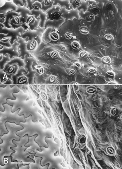
Figure 2.
LTSEM of TMV-induced HR lesions. A, Upper epidermis of TMV.GFP-infected N. edwardsonii leaves shortly after visible lesion development at 14 hpt. Turgid and collapsed epidermal cells can be clearly differentiated; at this time point guard cells retain turgor. B, A later stage of lesion development at 20 hpt with both epidermal and guard cells showing loss of turgor. Scale bar = 0.1 mm (A and B).

