Figure 6.
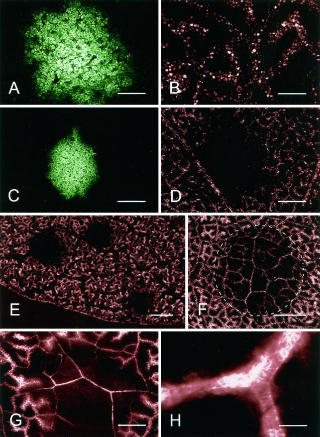
Transport of Texas Red through the xylem of TMV.GFP infection foci. CLSM images of TMV.GFP-infected and control-noninfected tissue 30 min after labeling of detached leaves with the fluorescent dye Texas Red. A, TMV.GFP infection focus at 10 hpt with the corresponding image of Texas Red shown in B. C, An infection focus at 11 hpt is shown with the corresponding Texas Red image in D showing a restriction of dye movement into the infected area of the leaf. E, Low-magnification image of a TMV.GFP-infected leaf at 11 hpt with areas of Texas Red exclusion corresponding to the position of three TMV.GFP infection foci. F, Texas Red in the xylem of a leaf following prevention of transpiration from both upper and lower epidermis after localized application of vacuum grease (encircled area). G, Resumed transport of Texas Red through a TMV.GFP infection focus at 20 hpt. H, High-magnification image of a TMV.GFP infection site 20 hpt showing that the fluorescent dye is restricted to the xylem elements. Scale bars = 0.5 mm (A and B); 1 mm (C and D); 4 mm (E); 2 mm (F); 0.5 mm (G); 25 μm (H).

