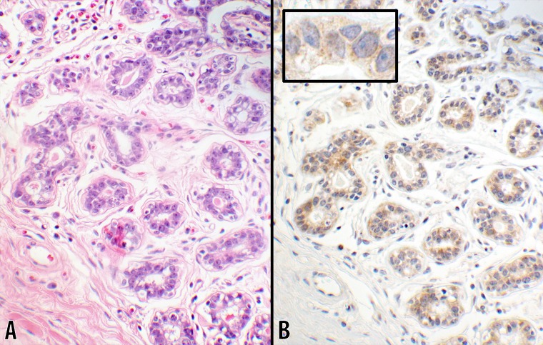
Fig 3. Score 2a in benign breast tissue (20x objective).
Photomicrographs of a normal (or benign) breast tissue showing the normal ductal structures with the adjacent interstitial tissue. B CTLA-4 stain with 1+ intensity showing a uniform light staining cytoplasmic granules in 100% of ductal cells. The luminal contents of the glands had a negative reaction. The inset is a portion of a ductal structure showing the sparsity of the cytoplasmic granules at a higher magnification (60x objective). A counterpart hematoxylin & eosin stain of the same tissue is shown in panel A (from #5; Table 1).

