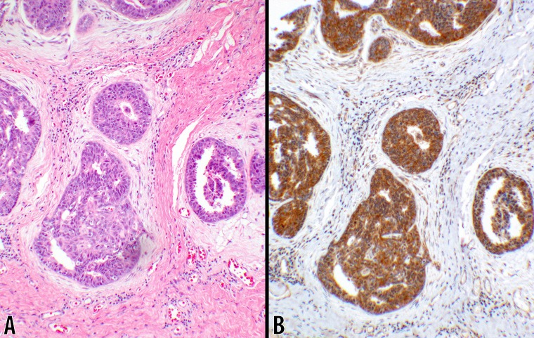
Fig 4. Score 2b in DCIS (10x objective).
An example of ductal carcinoma in-situ is shown where the neoplastic lesion is confined within the ductal structures. B CTLA-4 with 3+ intensity showing a uniform strong cytoplasmic stain in 100% of the neoplastic cells. A counterpart hematoxylin & eosin stain of the same tumor is shown in panel A (from #7; Table 2).

