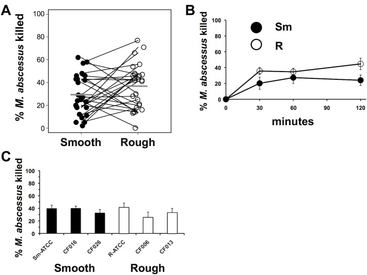
Fig 1. Neutrophil killing of M. abscessus.
(A) Neutrophils were incubated with smooth (closed circles) and rough (open circles) M. abscessus for 1h, and surviving mycobacteria were compared to the inoculum. Connecting lines represent results from the same donor neutrophils; n = 26. Mean values are depicted by the gray bars. (B) Time course of killing of M. abscessus morphotypes by neutrophils; n = 6. (C) M. abscessus clinical isolates were assayed for killing by neutrophils for 1 h; smooth morphotypes (closed bars); rough morphotypes (open bars); n = 4–10. None of the differences were significantly different for the Fig 1 data by paired t-test.

