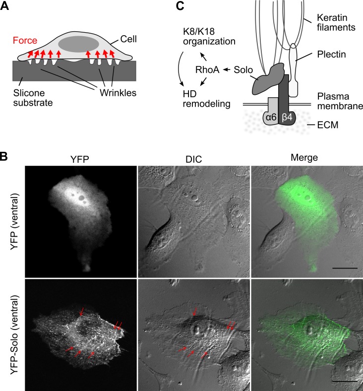
Fig 6. Localization of Solo at the sites of traction force generation and a model for the role of Solo in hemidesmosome remodeling.
(A) Schematic illustration of the side view of the cell on silicone substrates. Wrinkles appear on the substrate depending on the forces exerted by the cells. (B) Wrinkle formation assay. MCF10A cells were transfected with YFP or YFP-Solo, seeded on a thin Matrigel-coated silicone substrate, and cultured for 24 h. Ventral images of YFP (green) and phase-contrast images were acquired with a confocal microscopy. Red arrows indicate ventral localization of Solo, particularly along the wrinkles. Scale bar, 20 μm. (C) A model for Solo-mediated HD remodeling. Solo localizes at the site of force generation on the ventral surface of epithelial cells and promotes HD formation by activating RhoA signaling and reorganizing keratin networks.

