Figure 3.
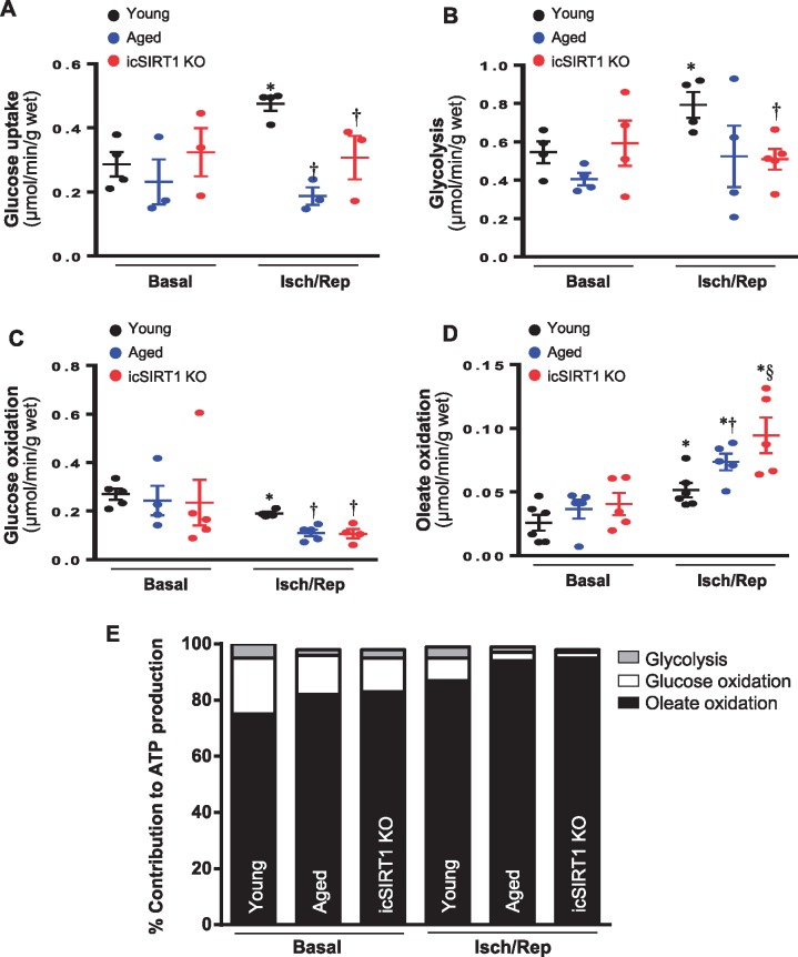
The mismatch substrate metabolism occurred in aged and icSIRT1 KO hearts. (A) d-[2-3 H]-glucose were used for determining the glucose uptake in young, aged, and icSIRT1 KO hearts under basal or ischaemia/reperfusion conditions. Values are means ± SEM, n = 3–4, *P < 0.05 vs. basal, respectively; †P < 0.05 vs. young I/R using 2-way ANOVA with Tukey’s post-test. (B) d-[5-3 H]-glucose were used in the heart perfusion system to measure glycolysis rate in young, aged, and icSIRT1 KO hearts under basal or ischaemia/reperfusion conditions. Values are means ± SEM, n = 4–5, *P < 0.05 vs. basal, respectively; †P < 0.05 vs. young I/R using 2-way ANOVA with Tukey’s post-test. (C) Glucose oxidation and (D) Oleate oxidation in the isolated working heart perfusion system. After balancing 20 min, isolated hearts were subjected to 10 min of ischaemia and 20 min of reperfusion. Glucose oxidation was analysed by measuring [14C] glucose metabolism into 14CO2. Oleate oxidation was measured by the incorporation of [9,10-3H2O] oleate into 3H2O. Values are means ± SEM, n = 4–6, *P < 0.05 vs. basal, respectively; †P < 0.05 vs. young I/R; §P < 0.05 vs. aged I/R using 2-way ANOVA with Tukey’s post-test. (E) Relative percentage of ATP production from glucose and oleate oxidation in young, aged, and icSIRT1 KO hearts.

