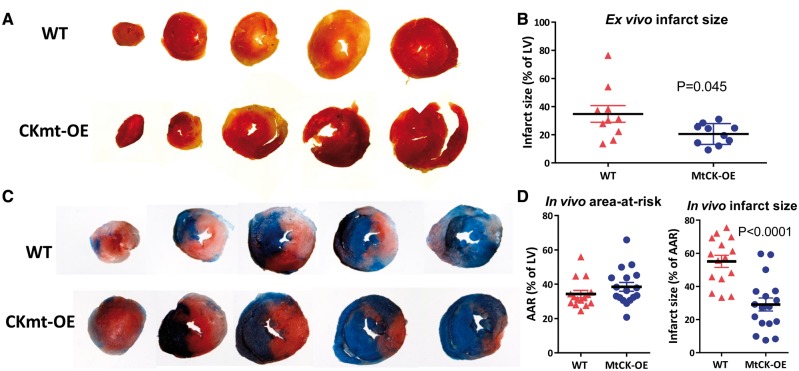
Figure 5.
Over-expression of MtCK reduces myocardial injury following ischaemia–reperfusion ex vivo and in vivo. At the end of experiments shown in Figure 4, perfused hearts were sliced and stained with triphenyltetrazolium chloride (TTC), which stains viable tissue red, while necrotic tissue appears pale in colour. (A) Shows representative sections spanning apex to base for WT and MtCK-OE hearts, with quantification of infarct size shown in (B) and comparison by two-tailed Welch’s t-test to reflect unequal variances. The analogous experiment was also performed in vivo with 45 min regional ischaemia and 24 h reperfusion. (C) Shows representative LV histological sections spanning apex to base. Myocardium that did not experience ischaemia appears blue, all other areas constitute the area-at-risk (AAR), while within this, necrotic tissue (infarct) appears pale or white. Quantification in (D) shows no difference in AAR, but a greatly reduced infarct size in hearts from MtCK-OE mice compared to WT mice using a two-tailed Student’s t-test (n = 17 and n = 15 females, respectively). Data are represented as mean ± SEM.

