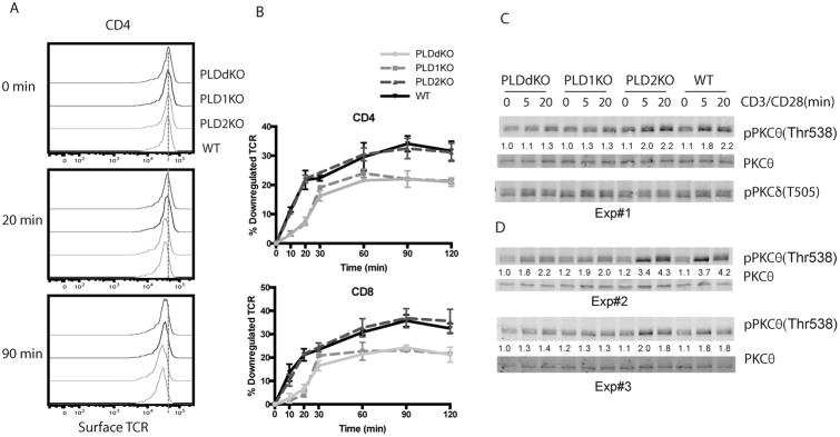
Figure 5. PLD-deficiency on TCR downregulation.
(A) Representative plots of surface TCR expression on splenic CD4+ T cells from PLDKO and WT mice before and after anti-CD3 and anti-CD28 stimulation. The dash line shows the mean MFI of CD3 before stimulation. (B) Quantitated TCR downregulation in CD4+ and CD8+ T cells. (C) PKCθ activation by Western blotting. Purified CD4+ T cells were stimulated with biotin-anti-CD3ε and biotin-anti-CD28, followed by streptavidin crosslinking for different time points. Cell lysates were analyzed by Western blotting with pPKCθ(Thr538), PKCθ, and anti-pPKCδ(Thr505). The relative intensities of pPKCθ was normalized by the intensity of pan-PKCθ. (D) PKCθ activation. Additional experiments showing that PLD1-deficiency impaired PKCθ activation.

