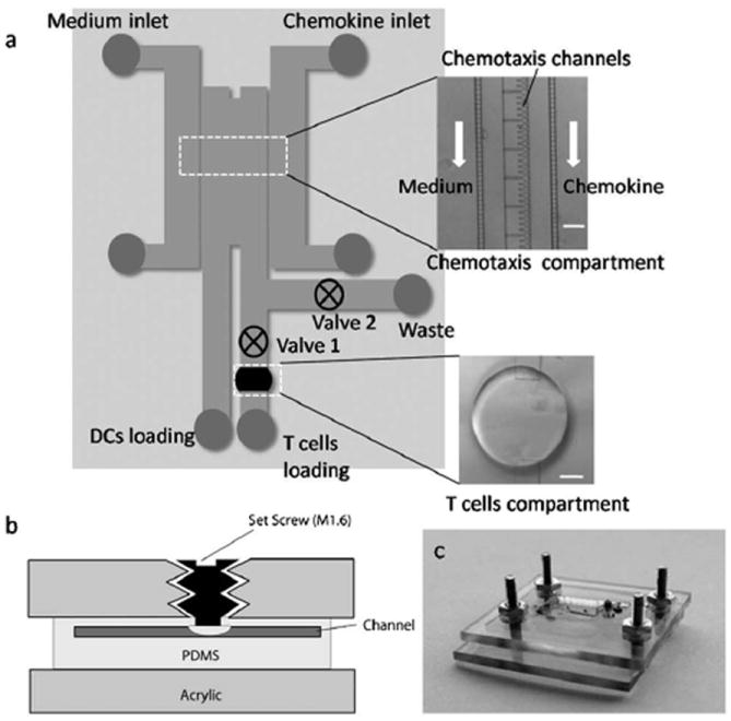
Fig. 1.

Schematic and images of microdevice. (a) Schematic of microdevice comprising of chemotaxis compartment connected to T cell compartment. The insets show micrographs of chemotaxis compartment and T cell compartment (scale bar: 100 μm). (b) Schematic illustrating basic operation of valve. (c) Image of the device mounted in the plastic manifold.
