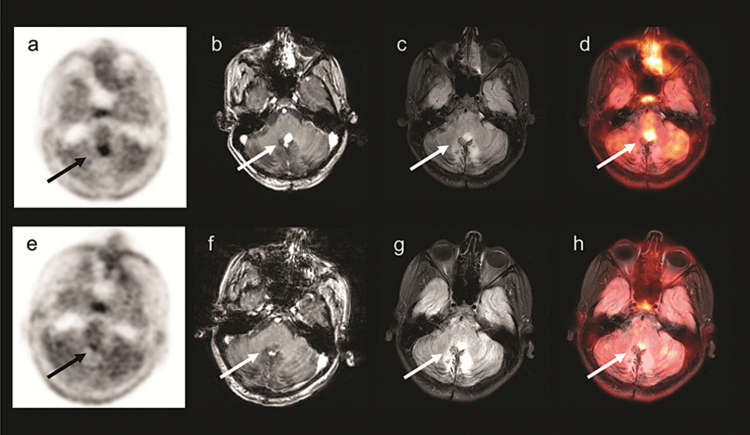
Fig. 2 .
A 12-year-old boy with cerebellar pilocytic astrocytoma (patient 2). Images prior to starting bevacizumab: (a)18F-FDOPA PET (b) T1-weighted MRI with contrast (c) Fluid-attenuated inversion recovery (FLAIR) MRI (d)18F-FDOPA PET/MRI. Images obtained 4 weeks following bevacizumab: (e)18F-FDOPA PET (f) T1-weighted MRI with contrast (g) FLAIR MRI (h)18F-FDOPA PET/MRI, showing near complete resolution of the tumor.

