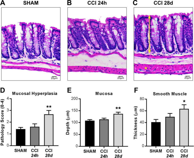
Figure 1. Histopathological evaluation in the colon at 24 hours and 28 days after TBI.

(A–C) Representative H&E staining and microscopy of colon sections from sham, 24 hours post-CCI, and 28 days post-CCI mice. (D) Pathology index for mucosal hyperplasia scored for severity and extent from 0 to 4. Morphometric analyses reveal (E) increased mucosal depth (double-headed arrrow) and (F) increased smooth muscle thickness (asterisk) in colons 28 days after CCI. Values (D–F) are means ± SEM; **P<0.01,*P<0.05, compared to shams; n=6-8.
