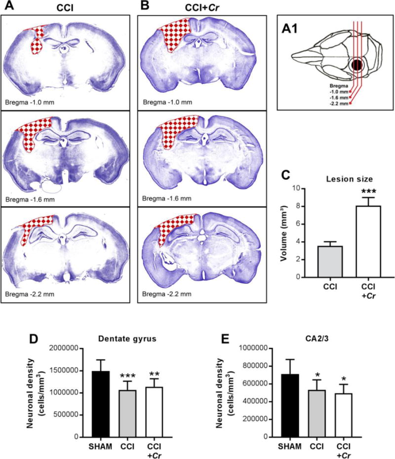
Figure 6. Enteric Cr infection during chronic TBI leads to greater cortical loss.

(A1–B) Schematic and representative cresyl violet-stained coronal sections showing CCI-induced lesions (red checkerboard fill) across injury site in brains of CCI and CCI+Cr mice. (C) Stereological analyses of lesion volume in whole brains on day 40 after CCI. Values are means ± SEM; ***P<0.001, compared to CCI; n=8–10. (D-E) Neuronal cell densities in the dentate gyrus and CA2/3 regions of the hippocampus. Values are means ± SEM; One-way ANOVA with Sidak correction for multiple comparisons *P<0.05, **P<0.01, ***P<0.001, compared to sham; n=8–10.
