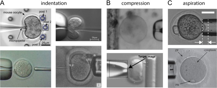
Figure 1.
Examples of microfluidic devices used to probe oocyte and embryo mechanical properties. The approaches to measure mechanics can be separated into three main categories. (A) Approaches involving indentation. The top left image is from Liu et al. (2012) and shows an oocyte being pushed against a group of flexible posts. The top right image is from Sun et al. (2003) and shows an oocyte deformed by a force-sensing microneedle. The bottom right image is from Green (1987) and shows an oocyte compressed by a quartz-fiber ‘poker’. The bottom left image is from Murayama et al. (2004) and shows the oocyte being probed by a MTS from the left side. (B) Approaches involving compression. The top image is from Abadie et al. (2014) and shows an oocyte about to be compressed between a micropipette and the edge of a floating platform. The bottom image is from Wacogne et al. (2008) and shows an oocyte being compressed between a micropipette and a flexible post (side view). The ‘target image’ text refers to the algorithm used to track the pipette displacement. (C) Approaches involving aspiration. The top image is from Yanez et al. (2016) and shows an embryo partially aspirated into a micropipette. The region between the arrows is the aspiration depth into the micropipette. The bottom image is from Khalilian et al. (2010b) and shows a portion of the ZP being aspirated into a micropipette.

