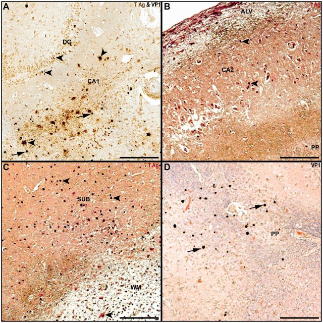
FIGURE 2.
Hippocampal gray and white matter JCV infection in patients with PML. (A) Granule cell neurons in the dentate gyrus and cells with astrocyte phenotype in CA1 express JCV T Ag (brown, arrowheads), whereas other glial cells in CA1 express JCV VP1 (blue-black, arrows). (B) Subpial demyelination (CNPase, brown) in alveus (ALV) and perforant path (PP) flank cells with phenotype of pyramidal neurons in CA2 expressing JCV Tag (red, arrowheads). (C) Multiple cells in the subiculum (SUB) express JCV T Ag (red, arrowheads) adjacent to an area of demyelinated white matter (CNPase, brown) containing an infected astrocyte (arrowhead). (D) Multiple glial cells in areas of demyelination in the perforant path (PP) express JCV VP1 (brown-black, filled arrows, with Luxol fast blue and hematoxylin and eosin counterstaining). Scale bars: 250 µm.

