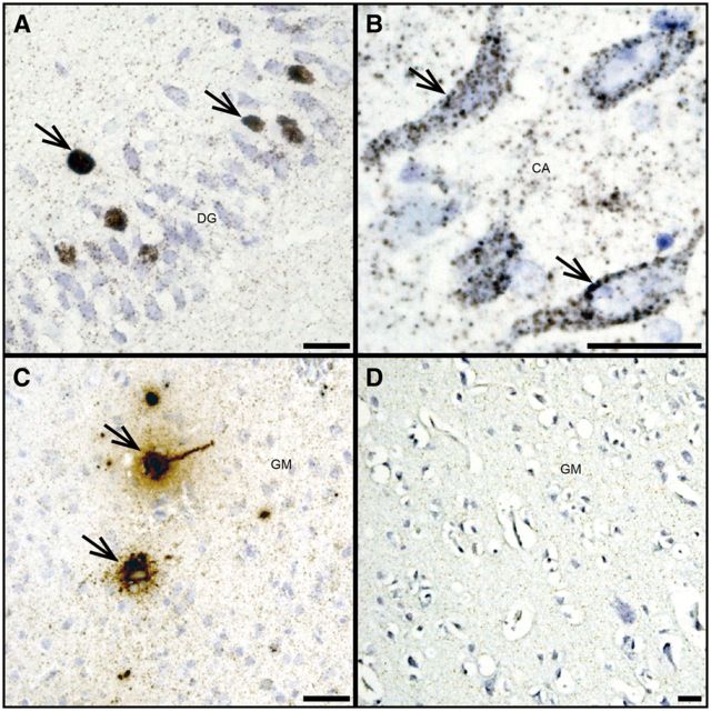
FIGURE 4.
JCV DNA is detected in hippocampal granule cell and pyramidal neurons. (A, B) JCV DNA (brown, arrows) is abundant in some granule cell neurons of the dentate gyrus of a patient with HIV/PML (A) and in some pyramidal neurons of the cornu ammonis of the same patient (brown, arrows) (B) . (C) High level of JCV DNA (brown, arrows) can be seen in pyramidal neuron of the hemispheric gray matter of a JCVE patient known to have almost exclusively JCV-infected neurons, demonstrated by presence of T Ag, VP1 by immunohistochemistry and viral particles by electron microscopy in a previous report ( 10 ), used as a positive control. (D) Absence of JCV DNA in the brain of an HIV-infected patient without PML used as a negative control. Scale bars: 25 µm.

