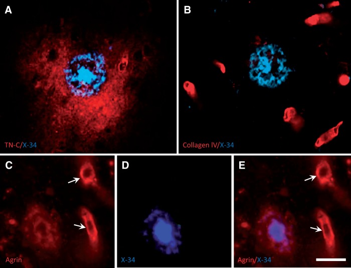
FIGURE 2.
High-magnification fluorescence microscopy images of frontal cortex tissue sections from an AD case, processed for dual labeling with X-34 and extracellular matrix proteins. (A) Typical X-34-labeled cored amyloid plaque (blue) is completely surrounded by larger diffuse TN-C deposit (red). (B) Collagen IV is detected exclusively in blood vessels (red) and is not seen in the X-34-labeled cored plaque (blue). (C–E) A proteoglycan agrin (red) is present in blood vessels (arrows) and in the periphery, but not the central core, of an X-34-positive Aβ plaque (blue). Scale bar: A – E = 30 µm.

