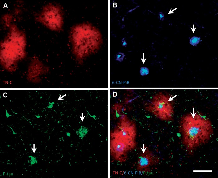
FIGURE 4.
Triple fluorescent labeling in a single section of frontal cortex from an AD case. Large TN-C deposits ( A , red) contain 6-CN-PiB-positive cored plaques ( B , blue) surrounded by clusters of phosphorylated-tau-ir dystrophic neurites ( C , green, arrows), whereas individual phosphorylated-tau-ir tangles show no TN-C immunofluorescence. Panel D shows merged fluorescence, with typical cored neuritic Aβ plaques surrounded by TN-C plaques (arrows in B – D ). Scale bar: A – D = 100 µm.

