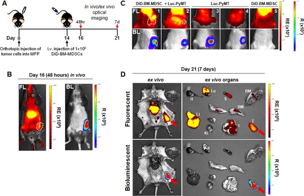
Fig 3. DiD-BM-MDSCs home to the primary breast tumor after adoptive transfer.
A) Schematic of treatment regimen for localization of DiD-BM-MDSCs in tumor-bearing mice. Mice were injected with 2.5x10⁵ Luc-PyMT cells into the MFP on day 0, and i.v. injected with 1x10⁶ DiD-BM-MDSCs on day 14. In vivo optical images were obtained at 48 hours (day 16) and 7 days (day 21) post-injection of DiD-BM-MDSCs. Ex vivo images were taken at 7 days post-injection (day 21). B) Representative in vivo FL and BL optical images on day 16; 48 hours after injection of DiD-BM-MDSCs. Tumors outlined by broken white line. C) Representative in vivo FL and BL optical images of mice from (A) on day 21; 7 days after injection of DiD-BM-MDSCs. Treatment groups include mice with Luc-PyMT tumors and adoptively transferred DiD-BM-MDSCs (mice 1–2; left), Luc-PyMT tumors alone (mice 3–4; middle) or DiD-BM-MDSCs alone (mouse 5; right). Tumors outlined by broken white line. D) Representative ex vivo FL and BL optical images of Luc-PyMT tumor-bearing mice (left) and individual organs (right) on day 21; 7 days after DiD-BM-MDSCs injection. Tumor indicated by red arrow. BM = bone marrow, H = heart, I = intestine, Ki = kidney, Li = liver, LN = lymph nodes, Lu = lungs, S = spleen, T = tumor. RE = Radiant Efficiency; R = Radiance.

