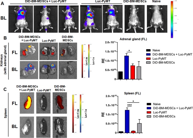
Fig 4. Homing of adoptively transferred DiD-BM-MDSCs to established metastases.
A) Representative in vivo images of Luc-PyMT metastases (BL signal) 3 weeks after i.c. injection into C57Bl/6 mice. B) Representative ex vivo images of DiD-BM-MDSC (FL signal; top panel) localization to adrenal gland metastases (BL signal; bottom panel). DiD-BM-MDSCs were injected (i.v.) into mice from A, and images acquired 2 weeks later. Quantification of radiant efficiency (RE) of FL-signal for the adrenal gland shown on the right. C) Representative images of spleens (left) and RE quantification (right) from treatment groups described in A and B. Naïve C57Bl/6 mice were used as controls. Data represented as mean ± SEM; *p<0.05; ***p<0.001; n = 3–5 mice for all groups.

