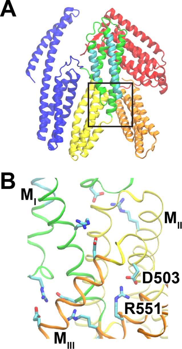
FIGURE 1:

α-Catenin structure. Structure from PDB ID 4IGG (Rangarajan and Izard, 2013). (A) α-Catenin consists of three major domains: the N-terminal (N, blue), modulatory (M), and C-terminal (C, red) domains. The M domain contains three four-helix bundles: MI (green/cyan), MII (yellow), and MIII (orange). MI contains the vinculin-binding site (cyan). The box indicates the region shown in B. (B) The salt bridge between D503 (MII domain) and R551 (MIII) is part of a salt-bridge network that stabilizes the structure of the M domain.
