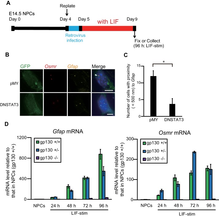
FIGURE 6:
Inhibition of JAK-STAT signaling impairs the clustering of Gfap and Osmr and down-regulates their transcription. (A) Scheme of experimental procedures. NPCs isolated from E14.5 mouse telencephalon were cultured and replated on day 4. The cells were infected with retroviruses that express EGFP alone (pMY) or together with DN-STAT3, and then cultured with LIF (50 ng/ml) for 4 d to induce astrocyte differentiation. (B) Projected images of double-labeled DNA FISH for Gfap (yellow) and Osmr (red) in LIF-stimulated cells that were infected with recombinant retroviruses engineered to express EGFP alone (pMY) or together with DN-STAT3. Virus-infected cells were stained with an anti-GFP antibody (green). Nuclei were counterstained with DAPI (blue). Scale bar = 5 μm. Arrowheads indicate clustering alleles. (C) Clustering frequencies determined using DNA FISH for Gfap and Osmr in LIF-stimulated cells that were infected with retroviruses expressing EGFP alone (pMY) or together with DN-STAT3. Data are presented as the means ± SEM from three biological replicates (n = 53–54). Student’s t test was performed. *p < 0.05. (D) Quantitative RT-PCR of Gfap and Osmr mRNA in NPCs derived from gp130 +/+, +/−, and −/− mice, alone and stimulated with LIF for different periods of time (LIF-stim). The results were normalized to Gapdh expression. Each graph represents the means (±SEM) relative to the amounts in NPCs derived from gp130 +/+ mice from at least three experiments.

