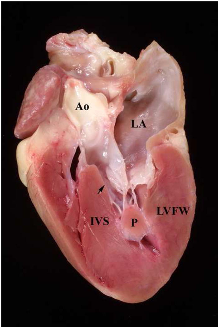
Figure 3.

Heart from a Maine Coon cat with HCM. The heart has been sectioned longitudinally. Note the thick interventricular septum (IVS), left ventricular free wall (LVFW), and papillary muscle (P). The arrow points to the narrowed left ventricular outflow tract. Ao = aorta, LA = left atrium
