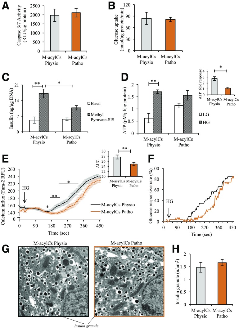
Figure 5.
Elevated M-acylCs impair glucose-stimulated ATP generation. We assessed caspase-3/7 activity (n = 3/group) (A), glucose uptake (n = 3/group) (B), methyl pyruvate–stimulated insulin secretion (SIS) assay (n = 5/group) (C), and steady-state ATP level (n = 3/group) (D) in intact murine islets treated with M-acylCs Physio and M-acylCs Patho concentrations in vitro. Calcium influx imaging was performed in dispersed murine islet cells treated with M-acylCs in vitro (n = 68 and n = 84 individual cells for M-acylCs Physio and M-acylCs Patho, respectively) and presented as average calcium influx, along with area under the curve (AUC) (E), and glucose responsive rate (F), where the cells with an increase in calcium influx >1.2 times of basal calcium were counted as glucose-responsive cells. Electron microscopic imaging was performed in intact murine islets treated with M-acylCs in vitro and is presented as a representative electron microscopic image (G) and insulin granule number (n = 10/group) (H). Scale bar, 500 nm. Data are presented as mean ± SEM. *P < 0.05, **P < 0.01. RFU, relative fluorescence units; RLU, relative luminescence units.

