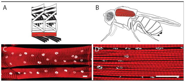
Figure 1. Larval and adult Drosophila muscles.
A) Schematic representation of the larval muscles. The Ventral Longitudinal (VL) muscles 3 (top) and 4 (bottom) are highlighted in red. B) Schematic representation of the adult muscles. The Dorsal Longitudinal Muscles (DLMs) that compose the major portion of the Indirect Flight Muscles are highlighted in red. C) Representative image of the 3rd instar larval VL muscles 3 and 4. D) Representative image of the DLMs where it is possible to observe multiple myofibrils and nuclei. C and D) Actin labeling in sarcomere, red; Nuclei, white. Scale bars, 25μm.

