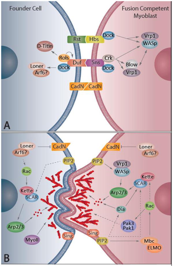
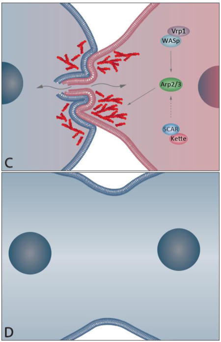
Figure 3. Schematic representation of myoblast fusion.

In the Drosophila embryo, fusion of an individual founder cell (FC) with multiple fusion competent myoblasts (FCMs) gives rise to a single multinucleated muscle cell; the number of FCM fusion events determines the number of nuclei in the mature muscle cell. A) Recognition and Adhesion: FC and FCM express different membrane proteins that allow the two cells to recognize and adhere to each other. When in contact, each cell initiates the fusion process by recruiting several proteins to the fusion site. B) Actin Focus Formation: Through the activity of the fusion machinery on both the FC and the FCM, actin (red) monomers are assembled into filaments and form a dense F-actin focus on the FCM side and a thin layer on the FC side. The FCM actin focus invades the FC with multiple finger-like protrusions. N-cadherin is removed from the membrane to allow for the next steps of fusion. C) Pore Formation. The formation of a fusion pore in the membrane occurs, allowing for the exchange of cytoplasm between both cells. The repression of the FCM transcriptional profile begins (FCM pink nucleus turns blue). D) Post Fusion. After one fusion event, the resulting cell has one additional nucleus that has the transcriptional profile of the FC (blue nuclei). At this moment, the cell either prepares for another round of fusion or stops fusing with FCMs.

