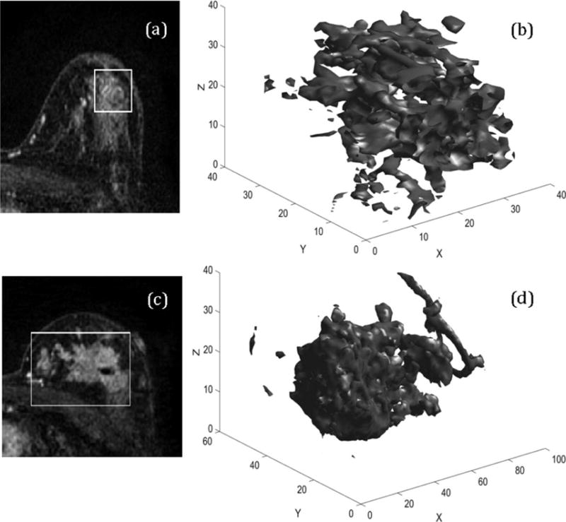
FIGURE 3.

3D representation of DCIS and DCIS upstaged to invasive carcinoma. a,c: Two central slices from the first postcontrast sequence of DCIS and DCIS upstaged to Invasive carcinoma using the radiologists annotation. b,d: The 3D renderings of the automatically segmented tumor. The values of the feature information measure of correlation1 are 0.1573 and 0.3919, respectively.
