
Extended data figure 10. Analysis of Tet2Δ/+;Flt3ITD and Tet2Δ/Δ;Flt3ITD leukemias.
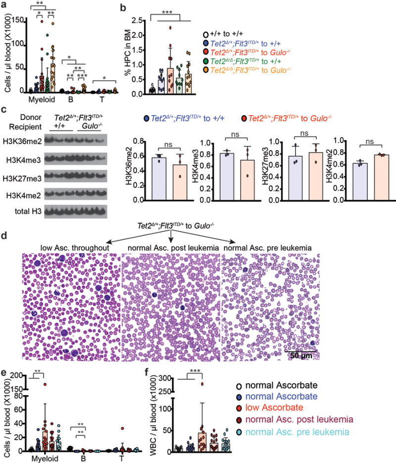
a-b, Analysis of recipient mice described in Fig. 4e–j. Statistical significance was assessed with one-way ANOVAs followed by Fisher’s LSD tests (b) or Kruskal-Wallis tests (a). c, Western blots with antibodies against the indicated histone modifications were performed using protein extracted from Tet2Δ/+;Flt3ITD/+ leukemia cells isolated by flow cytometry from wild type or ascorbate-depleted Gulo−/− transplant recipients (results are representative of 2 independent experiments). d, Diff-Quik stained blood smears from Gulo−/− recipients of Tet2Δ/+;Flt3ITD/+ cells fed with an ascorbate supplemented diet before and after the engraftment of leukemia cells (representative images from the experiments described in Fig. 5a and quantified in Fig. 5c). Cells with an immature blast-like morphology were more abundant in the blood of ascorbate-depleted Gulo−/− recipients as compared to ascorbate-fed Gulo−/− recipients. e-f, White blood cells in recipient mice from the experiment described in Figure 5a. The statistical significance of differences among treatments was assessed with Kruskal-Wallis tests (a, e, f) or a one-way ANOVA (b) or a two way ANOVA followed by Fisher’s LSD tests (c). All data represent mean±SD. Statistical significance was assessed with corrected for multiple comparisons by controlling the false discovery rate. (*p<0.05, **p<0.01, ***p<0.001).