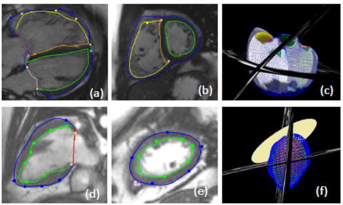
Figure 1.

Cardiac magnetic resonance images with epicardial and endocardial contours. a) long axis view b) short axis view and c) 3D biventricular model of a patient with tetralogy of Fallot. d) long axis view e) short axis view and f) 3D single ventricle model.
