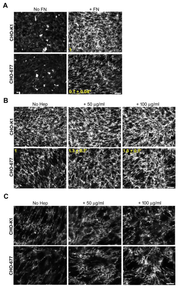
Figure 1. Heparin increases FN matrix in HS-deficient CHO cells.
Wild-type CHO-K1 and HS-deficient CHO-677 cells were grown for 3 days and, except for the “No FN” samples, exogenous FN was added at 25 μg/ml for the last 24 h. Samples were fixed and immunostained for FN, matrix was visualized by fluorescence microscopy, and fluorescence intensities were quantified. (A) Cells were grown with (+FN) or without FN (No FN). The fold change in mean fluorescence intensities of +FN samples for CHO-677 compared to CHO-K1 cells is shown in yellow (mean ± SE) (n=3, *p = 0.001). Scale bar = 50 μm. (B) CHO-K1 and CHO-677 cells were supplemented with FN alone (left), or together with 50 μg/ml (middle) or 100 μg/ml (right) heparin for 24 h. The fold change from the no heparin condition (No Hep) is noted for the CHO-677 cells (mean ± SE, n=3, *p < 0.05). Scale bar = 50 μm. (C) Higher magnification images of CHO-K1 and CHO-677 cells in (B) with FN and with or without heparin addition. Scale bar = 20 μm.

