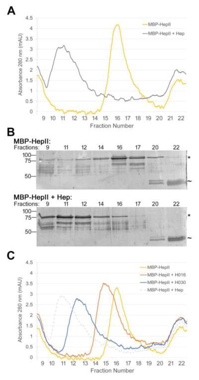
Figure 8. Heparin binds two or more HepII domains.
(A) 1 μM MBP-HepII alone (yellow) or preincubated with 10 μM unfractionated heparin (Hep) (gray) for 1 h was subjected to size exclusion chromatography. The elution profiles show A280 (milli-absorbance units, mAU) for fractions from the void volume (fraction 9, which contains protein aggregates) to ~ 20 kDa (fraction 22). MW standards eluted in fractions 9 (670 kDa), 14 (158 kDa), 21 (44 kDa), and 23 (17 kDa). (B) Fractions from (A) were analyzed by SDS-PAGE using 10% polyacrylamide SDS gels and silver staining. Molecular mass standards are indicated on the left. * indicates MBP-HepII, ~ indicates MBP at 42 kDa, which contaminates the MBP-HepII preparation. (C) Size exclusion chromatographs for 1 μM MBP-HepII alone (yellow) or incubated for 1 hr with 10 μM H030 (blue), H016 (orange), or unfractionated heparin (dotted gray). mAU is plotted versus fraction number. Elution profiles were confirmed by SDS-PAGE and silver staining (data not shown).

