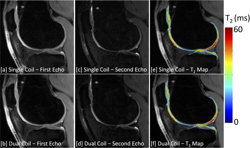
Figure 7.

3D DESS images of the [a,b] first echo, [c,d] second echo, and [e,f] the computed T2 relaxation time maps acquired using a single-coil [top row] and dual-coil [bottom row] configurations. Bilateral scans used a factor of 3 undersampling in the slice-encode (right-left) direction to acquire images of both knees simultaneously in similar scan times to traditional single-knee acquisitions.
