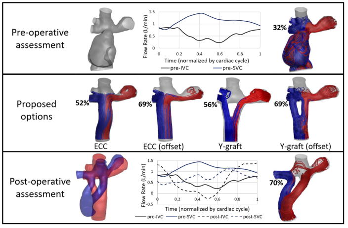
Fig. 7.
Representative surgical planning case. Only inlet waveforms are shown for clarity. Streamlines colored by outlet vessel (LPA=red, RPA=blue) HFD to the right lung is indicated by percentage. The Y-graft (offset) option was implemented in the patient. Post-operative assessment was conducted at a 2 month follow up visit. To compare anatomies, the proposed option (red) is overlaid with the actual post-operative anatomy (blue). (ECC: extracardiac conduit)

