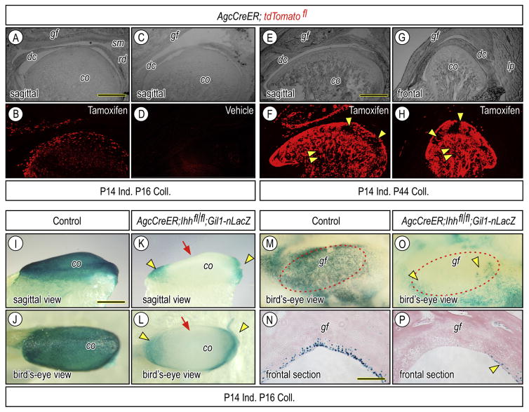
Figure 3.
Clarification of Cre recombinase activity and inactivation of hedgehog signal in postnatal TMJs of Agc-CreER; Ihh f/f;Gli1-nLacZ. Bright field (A, C, E, G) and corresponding fluorescence (B, D, F, H) images of parasagittal (A–F) and frontal (G–H) sections of TMJs from which Agc-CreER-tdTomato fl mice had been administrated tamoxifen once (A, B, E–H) or vehicle (C, D) at P14 and were sacrificed at P16 (A–D) or at P44 (E–H). Fluorescence images (B, F, H) show that reporter activity is detected in superficial cells, progenitors and chondrocytes of the articular cartilage of the glenoid fossa and condyle, but is absent from the articular disc (dc), retrodiscal tissue (rd) and synovial membrane (sm). Strong reporter activity is also detected in marrow cells (F, H: arrowheads). Whole mount LacZ staining (I–M, O) and frontal section (N, P) of condyles (I–L) and glenoid fossa (M–P) obtained from P16 -Agc-CreER; Ihh f/f;Gli1-nLacZ or control P16-Agc-CreER; Ihh f/f;Gli1-nLacZ that had been injected with tamoxifen (I–P) at P14. LacZ-positive cells resided at the anterior and posterior ends of the condylar articular cartilage (K, L, arrowheads) and at the medial, lateral, anterior and posterior edges of the fossa (O, P, arrowheads). Scale bars: 160 μm in A for A–D; 220 μm in E for E–H; 200 μm in I for I–M, O; 130 μm in N for N, P. gf, glenoid fossa; dc, articular disc; cd, mandibular condyle, lp, lateral pterygoid muscle.

