
Fig. 2.
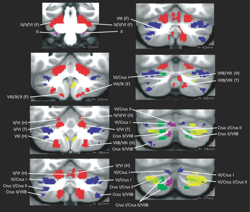
Summary of cerebellar activation for motor (red), working memory (blue), language (yellow), social processing (green) and emotion processing (magenta); coronal plane. (H) = hand, (F) = foot, (T) = tongue. Activation maps are thresholded at a voxel-level threshold of d>0.5. Only clusters >100 mmˆ3 are shown. Left is shown on the left.