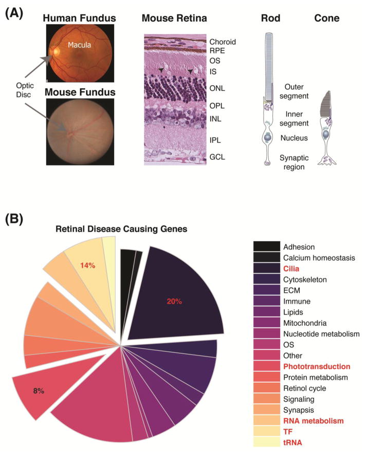
Figure 1.
Retinal structure and disease-causing genes. (A) Fundus images of a healthy human and mouse retinas, depicting the blood vessels, optic nerve head, and macula (in human). Histology of mouse retina, showing the laminar structure of the retina. Arrow heads point to the unique cone OS. A schematic representation of photoreceptors, highlighting the different morphological and functional domains of the cell. (B) Pie-chart of reported retinal degeneration genes (RetNet; https://sph.uth.edu/retnet), organized by the functional groups. The top three groups are indicated in red and pulled out of the pie. Abbreviations: RPE, retinal pigment epithelium; OS, outer segment; IS, inner segment; ONL, outer nuclear layer; OPL, outer plexiform layer; INL, inner nuclear layer; IPL, inner plexiform layer; GCL, ganglion cell layer; ECM, extracellular matrix.

