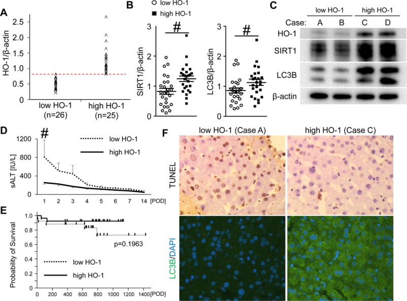
Figure 6. Increased post-transplant HO-1 levels correlate with attenuated hepatocellular damage and enhanced SIRT1 – LC3B pathway in human OLT.

(A) Bx samples collected from the fifty-one OLT patients were classified into low HO-1 (n=26) and high HO-1 (n=25) groups. (B) Western blot-assisted expression of SIRT1 and LC3B. Data shown in dot plots and bars indicate mean±SEM. # p<0.05 (Mann-Whitney U test). (C) Representative Western-blots assisted detection of HO-1, SIRT1 and LC3B (Case A/B: low HO-1 group, Case C/D: high HO-1 group) (D) sALT levels at postoperative day 1-14 (POD1-14) shown as mean±SEM. Dotted line indicates low HO-1, while the solid line high HO-1. # p<0.05 (Mann-Whitney U test). (E) The cumulative probability of OLT survival (Kaplan-Meier method). Dotted line indicates low HO-1, while the solid line high HO-1 groups (log-rank test). (F) Representative TUNEL and LC3B staining pattern in OLT.
