Fig. 1.

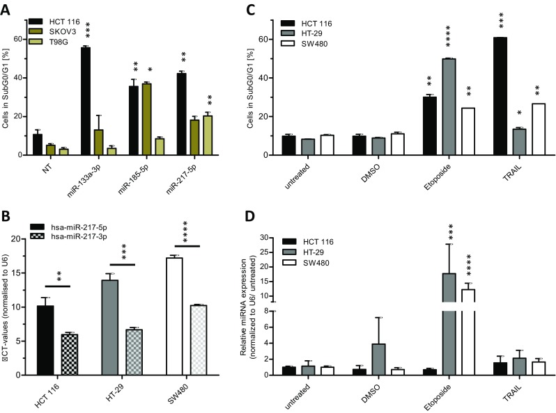
miR-217-5p expression is increased upon induction of the apoptosis. For validation screening HCT 116, SKOV3, and T98G cells were seeded 24 h before transfection with miRNA mimics (50 nM and 0.4 μl ScreenFect®A) or non-targeting siRNA (NT) control. Apoptosis rates 72 h after transfection were analyzed by Nicoletti staining followed by flow cytometric analysis (a). For miR-217-3p and -5p expression analysis total RNA was isolated from untreated HCT 116, HT-29 and SW480 cells and applied to cDNA synthesis followed by qRT-PCR. The expression analysis of both miR-217 strands was done by normalization to the CT value of U6 snRNA (b). For determination of miR-217-5p expression after induction of apoptosis, cells were seeded in 24 h prior treatment with Etoposide (25 μM), TRAIL (150 ng/ml, except HCT 116 with 80 ng/ml) or DMSO for additional 48 h. The apoptosis rates 48 h after treatment were analyzed by Nicoletti staining and flow cytometric analysis (c). The miRNA expression of miR-217-5p was normalized to the CT value of U6 snRNA and the untreated control (d). Statistical analyses for part (a, c and d) were performed by two-way ANOVA followed by Bonferroni post-test. Statistical differences for part B were tested using unpaired t-test. The treatments were compared to NT (a) or untreated cells (c and d) [n = 3 biological replicates; mean ± SD, *p < 0.05; **p < 0.01; ***p < 0.001; ****p < 0.0001]
