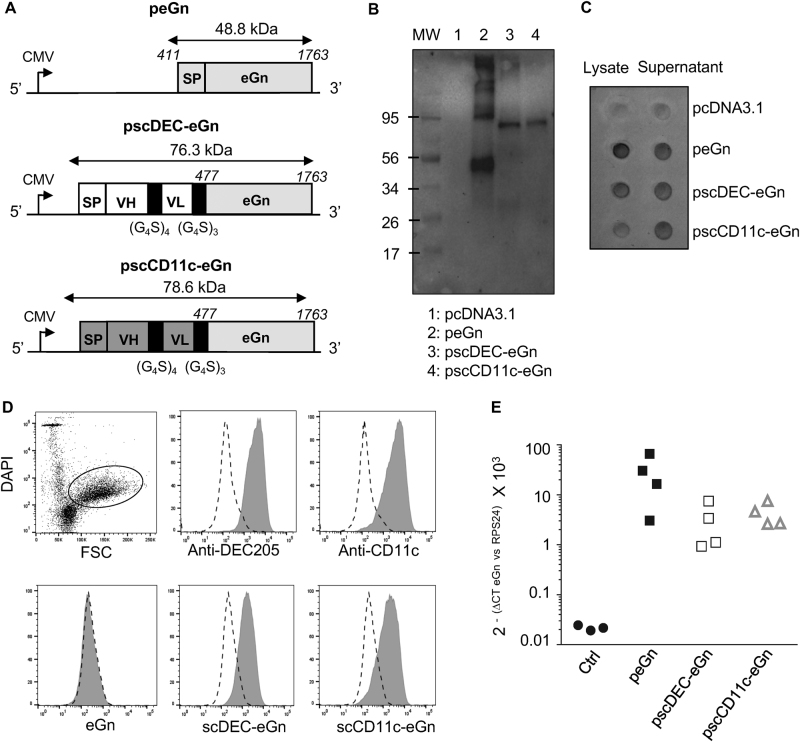
Fig. 2.
Design and expression of peGn, pscDEC-eGn and pscCD11c-eGn. a Schematic representation of peGn, pscDEC-eGn and pscCD11c-eGn. peGn includes the codon-optimized eGn sequence with its intrinsic signal peptide (SP, RVFV M segment from nt 411 to nt 1763) and cloned in the pcDNA4-V5His expression vector. pscDEC-eGn and pscCD11c include the signal peptide (SP) of the VH chain of the parental mAb sequence, the scFv (VH and VL) sequence and the nt 477 to nt 1763 portion of the codon-optimized eGn sequence. The VH and VL sequences are connected together with a (G4S)4 linker and the scFv and eGn sequences are connected with a (G4S)3 linker. These chimeric sequences were cloned in a pcDNA3.1 vector. b, c HEK293 cells were transfected with pcDNA3.1, peGn, pscDEC-eGn and pscCD11c-eGn. In (b), the cell lysates (10 µl) were separated by SDS-PAGE under reducing conditions and eGn was revealed with anti-RVFV HMAF followed by a HRP-GAM IgG. The predicted sizes of the expressed untargeted and chimeric eGn proteins are 48.8 kDa for untargeted eGn, 76.3 kDa for the scDEC-eGn protein and 78.6 kDa for the scCD11c-eGn protein. All samples were gathered from the same experiment and were processed in parallel (1 repeated experiment). In (c) the cell lysates (100 µl) and the concentrated cell supernatants (500 µl) gathered from the same experiment were spotted onto the same nitrocellulose membrane and the eGn protein was revealed as in (b) (1 repeated experiment). d Sheep skin lymph low-density cells were reacted with the parental anti-DEC205, anti-CD11c or isotype control mAbs (top panel) or with the concentrated supernatant from HEK293 cells transfected with peGn, pscDEC-eGn, pscCD11c-eGn and a negative control pcDNA3.1 (bottom panel). The staining of the DAPIneg FSChi cells, which include dominantly cDCs, is depicted (dark gray) compared to control (dash line). Bound mAbs were revealed with A488-DAM IgG and bound eGn with anti-RVFV HMAF followed by A488-DAM IgG. e Relative expression of peGn, pscDEC-eGn and pscCD11c-eGn in sheep skin. peGn, pscDEC-eGn and pscCD11c-eGn (100 µg) were injected intradermally in 100 µl saline in inner front leg sites of two sheep followed by SEP (532 V/cm). After 48 h, RNA was extracted from skin biopsies. eGn mRNA levels were measured using qRT-PCR, normalized with RPS24 ribosomal RNA and expressed as 2−ΔCT values (1 repeated experiment). Absence of residual plasmid in the RNA preparations was controlled using qPCR without reverse transcription

