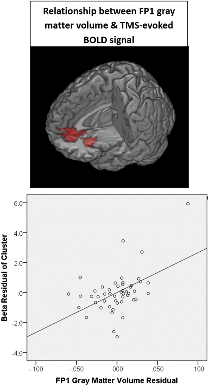
Figure 2.

Relationship between gray matter integrity and subcortical response to TMS. Using voxel-based morphometry, the gray matter volume at the site of stimulation and afferent targets (see Supplemental Figure S1 for ROIs) was isolated. As a group, there was a significant positive relationship between the gray matter volume in the cortical site of stimulation (FP1) and TMS-evoked BOLD signal in the anterior-cingulate, as well as the orbitofrontal cortex. Individuals with higher gray matter volume had a larger effect of TMS in these cortical afferent targets. A scatter plot shows the relationship between FP1 gray matter volume and cluster beta values after controlling for participant age, TMS pulses administered and scalp to cortex distance (B).
