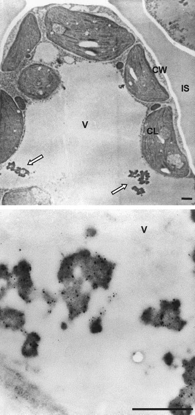
Figure 3.
SIgA/G forms protein body-like aggregates in the vacuole. Ultra-thin sections of transgenic tobacco leaves expressing SIgA/G (top panel) were incubated with anti-SC and anti-Ig antibodies (bottom panel). Detection was performed using secondary antibodies conjugated with 12 nm (for SC) and 6 nm (for Ig) colloidal gold. Arrows indicate the vacuolar protein body-like structures. CL, Chloroplast; CW, cell wall; IS, intercellular space; V, vacuole. Bars = 1 μm.

