Fig. 4.

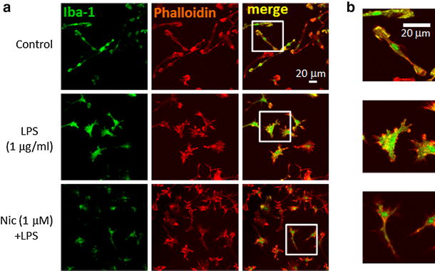
Effect of nicotine on LPS-induced morphological change of microglia. a Effects of LPS and nicotine on cellular morphology of microglia. Microglial cells were treated with LPS (1 μg/ml) for 24 h. Nicotine (1 μM) was pre-treated for 1 h before application of LPS. Immunofluorescence stained with anti-Iba-1 antibody (labeled with Alexa Fluor 488; green), and anti-phalloidin (anti-F-actin antibody) (labeled with Alexa Fluor 568; red) are shown. b Images in white squares in a are enlarged with different scale. More filopodia and membrane ruffling (actin polymerization) are shown in LPS-treated microglia
