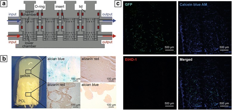
Fig. 3.

a Cross-sectional bioreactor schematic. b Macroscopic and histological analysis of engineered osteochondral interface. c Live/dead staining of capillary-like network formed by HUVECs in bone compartment (GFP, green = HUVEC; Calcein Blue AM, blue = live cells; EthD-1, red = dead cells). gelMA gelatin methacrylate, PCL poly(ε-caprolactone), GFP green fluorescent protein, EthD-1 ethidium homodimer-1
